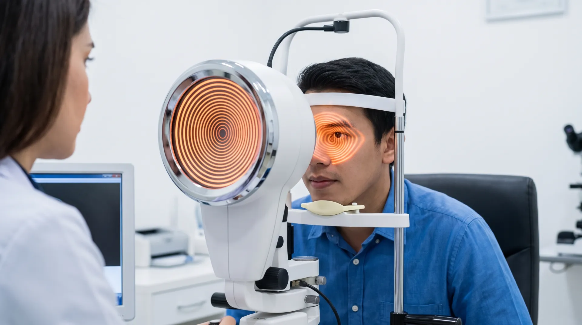
How Keratoconus Is Diagnosed: Advanced Corneal Imaging
Keratoconus is diagnosed using corneal tomography — specifically Scheimpflug imaging (Pentacam) — which maps both the front and back surfaces of the cornea. Unlike standard topography, which only images the front surface, Scheimpflug tomography detects keratoconus years earlier because the posterior cornea is where keratoconus shows first.
Medically Reviewed by Dr. Alexander Bonakdar, O.D.
What a Keratoconus Evaluation Includes
Corneal Tomography
Scheimpflug imaging (Pentacam) captures 25,000+ elevation data points on both corneal surfaces in approximately two seconds.
Corneal Topography
Placido-disc mapping of the anterior corneal surface curvature, providing SimK values and irregularity indices.
Pachymetry
Full corneal thickness mapping — not just a single central measurement — to identify focal thinning patterns.
Slit Lamp Examination
Biomicroscopic examination of the cornea for Vogt striae, Fleischer ring, corneal scarring, and hydrops signs.
Best-Corrected Visual Acuity
Manifest refraction to determine the best vision achievable with glasses, documenting the visual limitation keratoconus causes.
Specialty Lens Assessment
If scleral lenses are indicated, diagnostic trial lenses are selected based on tomographic elevation data.
What Is Scheimpflug / Pentacam Imaging?
The Pentacam uses a rotating Scheimpflug camera that captures cross-sectional images of the cornea as it rotates 360 degrees around the eye. In approximately two seconds, it generates a complete three-dimensional model of the cornea from 25,000 or more true elevation data points.
Unlike Placido topography (which only measures the front surface by analyzing reflected ring patterns), Scheimpflug tomography directly images both the anterior and posterior corneal surfaces, generating true elevation maps and a complete pachymetry (thickness) map of the entire cornea.
The test is non-contact, painless, and takes seconds. No drops or anesthesia are needed.
Key Measurements
- 25,000+true elevation data points captured per scan
- ~2 secscan time — non-contact and painless
- BAD-DBelin/Ambrosio Enhanced Ectasia Display — the gold-standard keratoconus index
- Bothsurfaces mapped — anterior and posterior cornea
Topography vs Tomography: Why It Matters
| Feature | Placido Topography | Scheimpflug Tomography (Pentacam) |
|---|---|---|
| Surfaces mapped | Front only | Front AND back |
| Early KC detection | Limited | Detects subclinical/forme fruste KC |
| Key index | SimK, I-S ratio | Belin/Ambrosio Enhanced (BAD-D) |
| Pachymetry | Central point only | Full corneal thickness map |
| Cross-linking candidacy | Insufficient alone | Required for documenting progression |
| Scleral lens fitting | Not used | Elevation data drives lens design |
Why the Posterior Surface Matters
Research has shown that keratoconus first manifests on the posterior (back) surface of the cornea before changes are visible on the anterior (front) surface. Standard Placido topography, which only maps the front surface, misses these early changes entirely.
Scheimpflug tomography detects this subclinical stage — called forme fruste keratoconus — when the condition can be treated with corneal cross-linking before significant vision loss occurs. This is why tomography, not topography, is the standard of care for keratoconus screening.
Early Detection Saves Vision
When keratoconus is caught at the subclinical stage, cross-linking can halt progression before the patient needs specialty contact lenses. Patients diagnosed at later stages may already have corneal scarring that limits their treatment options.
This is why we recommend keratoconus screening for anyone with a family history of the condition, and for all children of keratoconus patients by age 10.
Same-Week Keratoconus Testing Available
Advanced Scheimpflug imaging with same-week appointments. Insurance accepted.
Role in Cross-Linking Decisions
Insurance companies require serial corneal tomography to document keratoconus progression before approving corneal cross-linking. Scheimpflug imaging provides the objective, reproducible measurements needed to demonstrate that the cornea is getting steeper or thinner over time.
Without tomographic documentation of progression, cross-linking pre-authorization is typically denied. This is one reason why having baseline and follow-up scans at the same facility — using the same instrument — produces the most reliable comparison data.
Who Should Be Tested for Keratoconus?
Recommended Screening
- •Family history of keratoconus (first-degree relatives)
- •Children of keratoconus patients by age 10
- •Anyone considering LASIK or refractive surgery (pre-operative screening)
- •Frequent or unexplained changes in glasses prescription
- •Vision that is not correctable to 20/20 with glasses
- •Chronic eye rubbing
Insurance Coverage
Corneal topography and tomography are billed under CPT code 92025 as a medical diagnostic procedure. When performed for suspected or documented keratoconus (ICD-10 H18.6), the test is covered by some medical plans.
For complete insurance details, see our insurance coverage guide.
Insurance Plans We Accept
We work with most major vision and medical insurance providers to maximize your benefits.
Vision Plans
- VSP (Vision Service Plan)
- EyeMed
- Davis Vision
- Spectera
- Superior Vision
- Avesis
Medical Plans
- Medicare
- Blue Shield of California
- Anthem Blue Cross
- United Healthcare
- Cigna
- Aetna
- Tricare
*Medical insurance is often used for medically necessary contact lenses.
CareCredit Financing
Make keratoconus treatment affordable with CareCredit. We offer 0% interest financing for 6- and 12-month plans on scleral lens fittings and other treatments. Apply in minutes — no impact to your credit score to check eligibility.
Schedule Your Keratoconus Evaluation
Advanced Scheimpflug corneal imaging with same-week availability. We verify your insurance before your visit.
Or call: (949) 693-4900
Schedule Your Expert Consultation
Fill out the form and our team will reach out within 1 business day.
Last reviewed July 2026 by Dr. Alexander Bonakdar, OD